카페검색 본문
카페글 본문
-
Re: 극상근 석회화 건염... 2020 . 2025.10.13해당카페글 미리보기
cuff repair using side-to-side sutures. (A) Preoperative fat suppressed T2-weighted magnetic resonance imaging coronal view shows calcific deposits on the supraspinatus tendon within musculotendinous junction. (B) Arthroscopic findings...
-
무릎, 고관절 관절염 jama 2020년 논문 2024.10.29해당카페글 미리보기
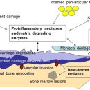
Figure 1B: Open in a new tab MRI (proton density, fat saturated) of right knee of 63 year old female. Coronal view on left and saggital view on right. Bone marrow lesions are identified with thin, solid white arrows on the coronal view...
-
Re: primary lateral sclerosis 2024 review 2025.04.16해당카페글 미리보기
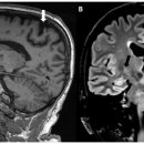
84,85] have been reported on T2- and FLAIR-weighted images, occasionally with a “wine glass” appearance in the coronal view at the diencephalic level [86], and the hypointensity of primary motor cortex (motor band sign) on...
-
Re: 럭비선수 햄스트링 파열 초음파 진단 2024!! 2025.07.10해당카페글 미리보기
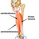
a) long-axis view, no bending of the CT and hematoma on the BFLH side of the CT; refer to the coronal MRI image; (b) short-axis view, hematoma on the BFLH side of the CT (white arrow), but not on the ST side. 4.5. Semitendinosus Muscle...
-
무릎 생체역학 구조해부학, 기능해부학 탐구!!! 2026.07.18해당카페글 미리보기
superior view (trochlear groove 위에서 본 patella contact zone)로 보여줍니다. PFJ mechanics의 dynamic load distribution을 직관적으로 이해할 수 있는 핵심 figure입니다. 1. Contact Pattern 20–30° flexion (가장 아래, white zone): Initial...