카페검색 본문
카페글 본문
-
Re: 극상근 석회화 건염... 2020 . 2025.10.13해당카페글 미리보기
cuff repair using side-to-side sutures. (A) Preoperative fat suppressed T2-weighted magnetic resonance imaging coronal view shows calcific deposits on the supraspinatus tendon within musculotendinous junction. (B) Arthroscopic findings...
-
무릎, 고관절 관절염 jama 2020년 논문 2024.10.29해당카페글 미리보기
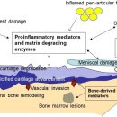
Figure 1B: Open in a new tab MRI (proton density, fat saturated) of right knee of 63 year old female. Coronal view on left and saggital view on right. Bone marrow lesions are identified with thin, solid white arrows on the coronal view...
-
Re: primary lateral sclerosis 2024 review 2025.04.16해당카페글 미리보기
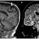
84,85] have been reported on T2- and FLAIR-weighted images, occasionally with a “wine glass” appearance in the coronal view at the diencephalic level [86], and the hypointensity of primary motor cortex (motor band sign) on...
-
Re: 럭비선수 햄스트링 파열 초음파 진단 2024!! 2025.07.10해당카페글 미리보기
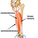
a) long-axis view, no bending of the CT and hematoma on the BFLH side of the CT; refer to the coronal MRI image; (b) short-axis view, hematoma on the BFLH side of the CT (white arrow), but not on the ST side. 4.5. Semitendinosus Muscle...
-
무릎 생체역학 구조해부학, 기능해부학 탐구!!! 2026.07.18해당카페글 미리보기
superior view (trochlear groove 위에서 본 patella contact zone)로 보여줍니다. PFJ mechanics의 dynamic load distribution을 직관적으로 이해할 수 있는 핵심 figure입니다. 1. Contact Pattern 20–30° flexion (가장 아래, white zone): Initial...
-
전침의 효과 3가지 관점 통시적 탐구(endorphin system, 항염증 system, 자율신경 전기자극), 통증 최신 기전까지 2026.05.12해당카페글 미리보기
물질이 이를 미세하게 조절 A. RVM의 위치 (시상면 Sagittal view) 뇌간의 아래쪽(medulla oblongata)에 위치한 RVM을 검은색...시스템의 핵심 출력 중추임을 보여줌. B. RVM의 횡단면 (Coronal section) RVM이 medulla의 복내측(ventromedial) 부분에...
-
Re: 디스크에 가해지는 부하에 대한 2026년 네이처 최신 논문 탐구 2026.03.05해당카페글 미리보기
Human coronal section from the back showing the three major lumbar spine muscles groups: the erector spinae, multifidus, and psoas major. D Transverse section showing relative positions of the intervertebral disc (including the annulus...
-
경추, 흉추, 어깨 통증의 생체역학적 통합!! 2026.05.12해당카페글 미리보기
sagittal view)으로 Latarjet과 Bristow 비교. (a) Latarjet: conjoint tendon이 subscapularis 하부를 돌아감 → 강한 sling effect (b) Bristow: subscapularis를 split해서 지나감 → sling 효과 약함 Figure 4 설명: 오른쪽 어깨 후방 관절경 시야...
-
shoulder 검사 입니다... (shouldr Axillary view 를 true Axillary 로 보는 방법 이 소개되었네요)..참고 하세요.. 2010.02.04해당카페글 미리보기
with the scapula flat against the cassette (as for the AP view in the plane of the scapula). The x-ray beam is centered...directed perpendicular to the cassette (45° to the coronal plane) except that it is angled 45° caudally. The beam...
-
Re: 고관절 골관절염 x ray 비정상 - no pain 많다!! 2024.10.28해당카페글 미리보기
profile view, the anterior JSW should always be larger than the posterior JSW [57, 64]. 정상 고관절 방사선 사진 정상 고관절의 방사선 사진에서는 일부 피질 불규칙성과 대퇴골 골화가 보일 수 있으며, 이는 OA 관련 구조적 뼈 변화와 혼동해서는...