카페검색 본문
카페글 본문
-
Re: 허리, 골반, 무릎, 발목 신경포착 탐구!! 2026.04.28해당카페글 미리보기
평균 34개월 (수개월~20년). 진단: 주로 임상 진단 (history + physical, Tinel’s sign 가능). 보조: Ultrasound (신경 비대, hypoechoic), MRI neurography (T2 high signal), NCS/EMG (감각 신경 속도 저하). Differential: Lumbar radiculopathy...
-
1990년대 이후 TrP(통증유발점) 기념비적 논문 탐구 2026.04.22해당카페글 미리보기
technology to visualize and characterize myofascial trigger points. Arch Phys Med Rehabil. 초음파로 MTrP를 hypoechoic nodule로 시각화 + stiffness 차이 확인. 비침습적 영상 진단의 시작(현재 2024 논문의 기반). 이전 연구들(특히 Shah et al.의...
-
thyroid adenoma 갑상선 선종 2026.04.11해당카페글 미리보기
Ultrasound as -solid -solitary -hypoechoic, or iso-echoic nodules -surrounded by a thin -complete peripheral halo thyroid adenoma is a benign (noncancerous), encapsulated tumor arising from follicular cells in the thyroid gland, often...
-
발 통증, 생체역학적 관점(Mechanical foot pain), 나이 관점의 탐구.. 2026.03.09해당카페글 미리보기
insertional microtears + degeneration). Diagnosis: Morning pain, first step pain, US에서 thickening (>4 mm) + hypoechoic change. Management: Stretching (calf, plantar fascia), orthotics (prefabricated/arch support), night splints, radial...
-
초음파 검사와 MRI 검사에서 암의 크기가 다를 수 있나요? 2025.11.24해당카페글 미리보기
있습니다. | 2. 병변의 특성 및 범위 측정 초음파의 과소평가 경향: 초음파는 종양의 **가장 단단하고 중심이 되는 부분(hypoechoic center)**만 측정하는 경향이 있어, 종양 주변의 침윤(invasion) 범위나 암세포가 퍼져있는 미세한 영역을 놓치기...
-
경추 불안정성으로 나타나는 문제들 2024 2025.11.13해당카페글 미리보기
symptoms with LCI and LUCI (24, 68). The optic nerve emerges from the posterior part of the globe and appears as a hypoechoic linear structure with a hyperechoic border (nerve sheath). The outer rim should be included in optic nerve...
-
Re: CIDP 만성 염증성 탈수초성 다발성 신경염증 진단과 치료 2020 2025.09.25해당카페글 미리보기
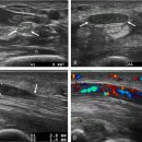
enlarged median nerve at the forearm with a CSA of 31.5 mm2 in a patient with CIDP. Some swollen fascicles and a hypoechoic structure can be depicted. Ultrasound stetting except focus are the same in both images CSA enlargements can...
-
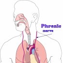
횡격막 근육의 기능부전 - 호흡불편함 phernic nerve(C3~5) 2025.08.27해당카페글 미리보기
layer structure, with two hyperechoic outer layers representing the pleural and peritoneal membranes, and the hypoechoic layer in the middle representing the diaphragmatic muscle [59]. Despite existing expert recommendations [60], there...
-
Re : 수근관 증후군 초음파 진단!! 2026.05.22해당카페글 미리보기
1, 3]. The smallest nerve structure that can be resolved by ultrasound is the nerve fascicle, which has a hypoechoic echo structure compared with the peri- and epineural connectives, which have a hyperechoic appearance and which are...
-
Re: Ultrasound-Guided Intervention in Cervical Spine 경추 초음파 진단 2025.07.15해당카페글 미리보기
of the brachial plexus (arrowheads) in the interscalene groove. These can appear as round or oval bundles with hypoechoic centers. C, carotid artery; C7 Tr, the 7th cervical transverse process. Click for larger image Figure 10 (A...