카페검색 본문
카페글 본문
-
Rare Cause of Exertional Angina 2024.01.03해당카페글 미리보기
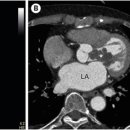
to our clinic with exertional chest pain for 2 weeks. Transthoracic echocardiography revealed a multi-lobulated hypoechoic mass occupying the left ventricular (LV) outflow tract with dynamic systolic protrusion into the aortic root (Fig...
-
견갑골 움직일 때 '딸각'거리는 소리 snapping scapular ... 2024.10.22해당카페글 미리보기
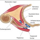
its mass effect, creating the appearance of scapular winging, together with crackles, and altering the scapulothoracic movement. It can also cause neurovascular compression, fractures, inflammation of the bursa, or malignant...
-
Rt. foot hemangioma^ 2012.07.16해당카페글 미리보기
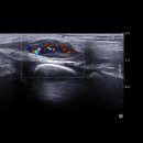
Palpable 상태 임. [Reading] Rt. foot US: Rt. midfoot 의 medial aspect에 navicular bone level에 lobulated contour의 hypoechoic mass lesion이 있음. 크기는 0.6x0.8x1.8cm이며 peripheral portion은 isoechoic thickening을 보이고 central portion...
-
암이 자라지 않는다네요... 2010.03.03해당카페글 미리보기
bladder (2.3 *1.8*1.0 cm) ; bladder cancer .TCC ,highly suspected . 2.known nephrectomy state,left . 3.a focal hypoechoic mass-mimic densit with central echogenic portion ,RK (r/o ,renal tumor mass ). 4. an echogenic nodule ,right liver...
-
Retroperitoneal extra-adrenal paraganglioma가 있는 10마리의 개에서 영상 features 2022.02.13해당카페글 미리보기
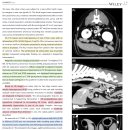
하며 irregular한 margin으로 확인됩니다. *Figure 5 : 9살령의 CM Boxer의 US 영상입니다. - (A) : Lobulated hypoechoic mass가 CdVC로 invasion하는 것이 확인됩니다. - (B) : 해당 mass는 좌측 부신의 인근에 존재합니다. - (C) : 반대편의 정상적인...
-
Imaging Findings of Chest Wall Lesions on Breast Sonography 2016.05.10해당카페글 미리보기
of a pyogenic abscess or simply an enlarging mass.5 When an abscess develops, sonographically, a heterogeneous hypoechoic mass with irregular borders appears in the chest wall (Figure 4⇓). Sonography is of particular value for detecting...
-
Renal Abscess 2016.05.02해당카페글 미리보기
AT III and fibrinogen. Ultrasonography: Sonographic signs for a renal abscess in renal ultrasound are a hypoechoic mass within the renal capsule, which may have inclusion of air (echogenic reflex with dorsal shadowing). Doppler...
-
(갑상선암) 세침흡입검사 결과 문의 2012.07.05해당카페글 미리보기
검사 이상, Thyroid gland의 left lobe에 1.0*0.9.0.8cm 크기의 irregular, microlobulated, and hypoechoic mass가 보임. Suspiciou malignant mass임. 이 mass에 대하여 US guided FNA를 2회 시행하였음. Mass는 thyroid capsule에 protruding하는 양상...
-
삼성서울병원 로봇수술 반절제 예정 2018.01.21해당카페글 미리보기
재판독 결과 5 (high suspicion), 12월 시행한 초음파상 기도랑 인접해있는 right lobe inferior 쪽으로 8mm irregular hypoechoic mass 나왔습니다. 양측 lateral neck에 의미있게 커진 lymph node는 보이지 않는다고 나왔는데 제가 걱정되는건 암위치가...
-
꼭 답변 부탁드릴게요 2009.12.21해당카페글 미리보기
Both thyroid glands reveal normal echopattern. Ill defined hypoechoic mass is seen in right lobe, which should be excluded of...
