카페검색 본문
카페글 본문
-
폐암2기? 4기? 교수님들 의견이 다릅니다. 어떻게 해야할까요? 조언부탁드립니다. 2017.02.19해당카페글 미리보기
and another smaller satellite nodule in the same lobe combined with metastatic lymph node at the right hilar and interlobar, the posterior aspect of the bronchus inermedius, indicating T3 N1 non-small cell lung cancer. - 우하방엽에 1.8cm...
-
판독 결과가 나왔는데 해석 좀 해주세요,,, 2005.07.12해당카페글 미리보기
consolidation 및 both lungs에 random distribution 을 보이는 numerous tiny nodule 및 cystic lesino이 있음 Right interlobar and hilar, left left, subcarinal , both paratracheal , left supraclavicular nodal station에 enlarged lymph node가...
-
청동기님 소견소 해석좀 해주세요~~ 2012.05.17해당카페글 미리보기
CT Chest routine (CE) [Finding] Breast cancer로 op. 시행받은 환자이다. Right middle lobe와 right lower lobe에 interlobar septal thickening이 증가하였으며 이는 lymphangitic metastasis의 aggravation과 postobstructive lymphedema의 가능성을...
-
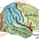
전두엽 2019.06.15해당카페글 미리보기
inferior Fusiform gyrus 37 Medial temporal lobe 27 28 34 35 36 Inferior temporal gyrus 20 Inferior temporal sulcus Interlobar sulci/fissures Superolateral Central (frontal+parietal) Lateral (frontal+parietal+temporal) Parieto-occipital...
-
X-ray 부위별 structures shown 2016.07.08해당카페글 미리보기
lordotic projection 전후 방향 척추전만 투상 . structures shown: used to demonstrate the lung apices and interlobar effusions 폐 정점 및 폐엽사이 액체를 설명하기 위하여 사용하는 AnteroPosterior axial projection 전후 방향 축 투상...
-
갑상선 수술후 PET-CT 판독결과 해석 부탁드려요 2013.11.29해당카페글 미리보기
in LUL of lung. - Rec) Follow-up to exclude non-FGD avid malignancy. 3. increased FDG uptake in bialteral hilar/interlobar and subcarinal arears, r/o reactive lymphadenopathy 4. Otherwise, no abnormal focal hypermetabolic lesion...
-
결과지좀 해석 부탁드리겠습니다^^ 2017.01.02해당카페글 미리보기
Benign lesion의 가능성이 높으나 follow-up을 통해 확인할 것을 권장함. 양측 pulmonary hilar lymph nodes, 양측 interlobar lymph nodes에 mild하게 증가된 FDG 섭취가 관찰됨. 이 부위에 high-attenuation이 동반되어 있음. Benign reactive change의...
-
내용 설명 해주실수 있나요...( 의학용어 ) 2004.02.01해당카페글 미리보기
기타 폐실질에 특이소견 없슴. 중심부 기도 및 폐혈관에 특이소견 없슴. 양측 늑막(강)에 이상소견 관찰되지 않음. Rt. interlobar, hilar, sub and precarinal, Lt. tracheobronchial and AP window region에 다수의 경계선상의 임파절들이 관찰됨. 스캔...
-
CT를 통해 흉수가 종양성인지, 염증성인지 구분할 수 있을 것인가? mesothelioma 감별의 impression 2022.05.20해당카페글 미리보기
neoplasia 진단에 impression을 더 줄 수 있는 CT상 특징으로는 Pleural thickening이 1cm 이상, nodular thickening, interlobar thickening, thoracic volume contraction 등이 있었으며, 본 연구의 목적은 더 큰 모집단을 통해 pleural malignant...
-
[인체 생리학] The Kidneys 2007.03.11해당카페글 미리보기
구조 : vascular elements와 tubular elements로 구성 ① Vascular component (nephron의 혈관) [renal artery → segmental → interlobar → arcuate → interlobular arteries → afferent arterioles] ⅰ) Afferent arteriole nephron에 도달되는 혈액...

