카페검색 본문
카페글 본문
-
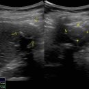
Hepatic isoechoic lesion with echogenic band?? (궁금합니다.) 2013.02.15해당카페글 미리보기
66세 여성으로 복부 초음파 검사중 우연히 발견된 case입니다. 결과가 아직 나오질 않았는데 궁금하네요. 개인적으로 complicated cyst일거라고 생각되는데?? 여러분들의 생각은 어떤지 듣고 싶네요. 수고하세요.
-
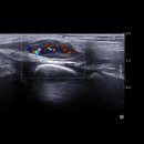
갑상선 유두암 수술 후) 초음파 결과지 해석좀 자세히 해주세요(수정) 2012.08.03해당카페글 미리보기
결과라고 썼을까요?) TSH 0.07 재검한 결과 ulu/ml TG < 0.1 ng/ml TG-Ab <20 U/ml About 0.64*0.64*1.4cm sized isoechoic soft tissue lesion at Rt.op. bed. --> possible remnant thyroid tissue. Lt.op.bed : negative No significant cervial LAP...
-
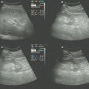
오늘 받은 방사선과 소견서입니다.. 판독좀 부탁드릴께요.. 2008.11.18해당카페글 미리보기
한 증가된 echo를 보이며, right lobe 에 각각 3.8 x 3.9x4.7cm , 4.5x4.2x3.9 cm size의 two large heterogeneous isoechoic round mass lesion 과 주위의 thin low echoic rim이 보임 Left lobe에도 1x1.3cm size의 small slight hyperechoic lesion이...
-
Rt. foot hemangioma^ 2012.07.16해당카페글 미리보기
foot US: Rt. midfoot 의 medial aspect에 navicular bone level에 lobulated contour의 hypoechoic mass lesion이 있음. 크기는 0.6x0.8x1.8cm이며 peripheral portion은 isoechoic thickening을 보이고 central portion은 anechogenicity를 보임...
-
Breast Ultrasonography 2016.04.25해당카페글 미리보기
almost isoechoic, as in the previous 2 cases, but in this patient significant tubular dilatation exists. The effect of ultrasound focusing. The focal zone, indicated by the white carrot and red arrow is appropriate in the left pane...
-
초음파와 조직검사결과지 해석좀 부탁드립니다. 2012.06.29해당카페글 미리보기
hypoechoic lesion in 9:30 of L breast. Still noted focal asymmetric parenchyma in 10:00 of R breast. Focal hypoechoic parenchymal change is noted in 6:00 of L breast. Ductectatic change in both breasts are noted. Thyroid USG: Right...
-
갑상선 결절 0.3센티인데.. 이정도 크기는 모양이나 석회화소견보여도 조직검사 안하는지 궁금해요. 2013.09.13해당카페글 미리보기
전 어떻게 하는게 좋을지.. 소견 부탁드려요.. 검사결과 내용 같이 첨부해봐요. both thyroids에 0.5cm이하의 hypo-to isoechoic nodules 여러 개 보임. left 0.3cm hypoechoic nodule은 indeterminate lesion이나 aspiration하기에는 크기가 작아 시행...
-
초음파 의학용어... 2004.09.18해당카페글 미리보기
echogenic lesion 에코 발생 병소, 메아리가 있는 병소 echogenic mass 에코 발생 종괴, 메아리가 있는 종괴 echogenic spot 에코 발생(메아리가 있는) 점 echogenicity 에코 발생도, 메아리 발생도 echogram 초음파 (영)상, 메아리 영상 echography...
-
Lt. renal mass like lesion 2005.05.11해당카페글 미리보기
focal lesion. GB and biliary trees are not dilated. Spleen, pancreas and Rt kidney are not remarkable. Somewhat isoechoic mass like lesion is seen in Lt kidney, probably normal anatomic variation. Imp; Diffuse liver disease. adv ; follow up
-
M/40 FNA was done. 2011.07.13해당카페글 미리보기
mixed echogenic round nodule containing intermal protruding mass. 2번 : Rt 2.0 by 2.0 cm sized isoechoic nodule mixed with cystic lesion 3번: Rt. 0.7 * 0.8 cm sized round isoechoic nodule * hyper, isoechogenicity는 갑상선 조지과 비교해서...


