카페검색 본문
카페글 본문
-
부인과 자기공명영상[pelvis magnetic resonance imaging, pelvis MRI] 2023.12.30해당카페글 미리보기
부인과 자기공명영상[pelvis magnetic resonance imaging, pelvis MRI] 한 줄 설명 초전도 자석과 고주파 및 컴퓨터를 이용하여 부인과 질환을 평가하기 위한 검사 진료과 ?해당 과를 클릭 하면진료과로 바로 연결됩니다. 영상의학과 진단 자궁근종, 자궁...
-
MRI : Bony Pelvis, Knee, Shoulder & Elbow - 기초가 중요하다는 걸 보여주는 자료 2012.03.08해당카페글 미리보기
구조와 Pathology에 대해서 완전하게 알지 않으면 MRI, 초음파 등의 진단도구를 이용할 수 없다. panic bird.. Musculoskeletal MRI. pelvis and knee.pdf INTRODUCTION Imaging of the bones, joints, muscles, and cartilage represents the radiologic...
-
수텐 치료 중 뼈 전이 의심 2026.06.27해당카페글 미리보기
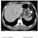
WO CONTRAST CLINICAL INDICATION: GIST peritoneal carcinomatosis. TECHNIQUE: Multisequence, multiplanar MRI of the abdomen and pelvis was performed with and without intravenous contrast. ESRC.2.13.3 COMPARISON: MR 3/24/2026. FINDINGS...
-
목회자가 병원 심방 시 꼭 알아야 할 의학 용어들 2026.06.14해당카페글 미리보기
Imaging(MRI): 자기공명영상 – 자기장과 고주파를 사용하여 신체 내 장기 및 구조의 상세한 이미지를 생성하는 진단 절차 Musculoskeletal: 근골격 – 신체의 지지 구조인 뼈, 인대, 관절 및 근육 Myocardial Infarction: 심근경색 – 동맥이 막히거나...
-
meralgia paresthetica 치료가 어렵지 않은데!! 2026.01.03해당카페글 미리보기
the pelvis and lateral to the psoas muscle, it crosses the iliacus before passing beneath the inguinal ligament. The LFCN then traverses medial to the anterior superior iliac spine (ASIS), ultimately piercing the fascia lata before...
-
MR neurography라는 MRI진단에 대한 논문, MRI의 진화 2024.10.14해당카페글 미리보기
한다. mri 사진보다 더 중요한 것은 neurologic exam에 의한 symptom, sign임을 MR neurography and muscle MR imaging for image diagnosis of disorders affecting the peripheral nerves and musculature MR neurography and muscle MR imaging 말초...
-
허리 MRI사진(횡단면)에서 구조물 확인 힘들어죠^^ 여기 그림 짱인 자료- Atlas of Sectional Anatomy(Abdomen and Pelvis) 2014.04.04해당카페글 미리보기
Atlas of Sectional Anatomy(Abdomen and Pelvis).pdf
-
CT, PET-CT, MRI 판독결과 해석 좀 부탁드립니다. 2010.02.25해당카페글 미리보기
and pelvis was obtained. the obtained images were reconstructed into axial, sagittal, and coronal planes. ct scan was performed from the neck to the proximal thighs after pet scan with contrast (omnipaque 300 inj, 150ml, 1 vial...
-
복잡한 고관절 통증 원인 찾기 2024.11.08해당카페글 미리보기
requires MRI or computed tomography.12,21 OSTEOARTHRITIS In older adults, osteoarthritis of the femoroacetabular joint is the most common cause of anterior hip pain.3 It can lead to significant morbidity and decrease in physical activity...
-
지독한 골반통증의 원인 '천장관절 증후군' 2024.11.08해당카페글 미리보기
모호합니다. MRI는 천장관절염을 발견하는 가장 민감한 영상 기술입니다. 천장관절 주변의 골수 부종과 염증을 확실하게 드러낼 수 있는 유일한 영상 기법이며 미란과 강직증을 입증하는 데 저선량 CT와 비슷합니다.21 후향적으로 요통의 다양한 원인으로...
![부인과 자기공명영상[pelvis magnetic resonance imaging, pelvis MRI] 이미지](https://search1.kakaocdn.net/argon/130x130_85_c/7il75sG5YQK)