카페검색 본문
카페글 본문
-
[조직검사지] 해석 부탁드려요 2007.11.20해당카페글 미리보기
x17cm with overlying ellpsoid skin(13x5cm). On serial sections of the breast parenchyma shows a lacting breast. A spiculated rubbery solid mass.measuring1.3x1cm is noted in the outer lower of quadrant. A tiny tissue defect site is noted...
-
갑상선암의 초음파 소견 2011.05.15해당카페글 미리보기
경우는 거의 대부분 양성 결절이고 악성의 가능성은 매우 낮으므로 주의를 요합니다. 결절의 경계가 불규칙하고 침상경계(spiculated margin) 모양을 보이는 경우도 악성결절의 가능성이 높습니다. 그러나 예민도는 낮습니다. <침상경계(spiculated margin...
-
오기근 지음 출판사 일조각 | 2008.02.20 2011.12.06해당카페글 미리보기
Calcifications case 36 Ductal Carcinoma in situ: Microcalcifications case 37 Infiltrating Ductal Carcinoma: Spiculated Mass case 38 Gynecomastia case 39 Asymmetry case 40 Normal Galactogram case 41 Quality Control 2단계 case 42 Focal...
-
갑상선 조직+세포검사 )) 병리검사 결과보고서 등 해독 요망 2009.11.03해당카페글 미리보기
Left lobe : neagtive - Isthmus : 1 nodule 1) IS1:rt, 11 mm, capsular (<50%), solid, hypoecho, microca++, spiculated / suspicous malignancy 3. Lymph nodes : No significant cervical lymphadenopathy 1) Central neck : An indeterminate small...
-
갑상선 혹에 대해 알아봅시다 2016.05.30해당카페글 미리보기
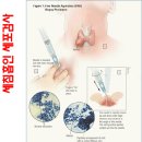
다음과 같습니다. 1. 위아래가 긴 모양 (taller than wider) 2. 미세 및 거대 석회화 3. 현저한 저에코 고형 결절 4. 침상(spiculated) 혹은 불규칙한 경계 갑상선 세침흡인검사 초음파를 보고 악성이 의심되는 소견이 보이면 갑상선 세침흡인세포검사를...
-
갑상선 검사 결과 문의드립니다.. 2012.09.02해당카페글 미리보기
해주셔서 감사드립니다.^^ 60대이신 저의 아버지 갑상선 결과 문의드립니다. 검사결과 1차초음파 : 1. Solid taller spiculated markedly hypoechoic nodule, RMP(0.31cm) -suspiciously malignant nodule ===US-guided FNAB was done 2.Solid,round,ill...
-
머리뼈와 척추에 발생한 osteosarcoma의 MRI 상 특징 2022.08.13해당카페글 미리보기
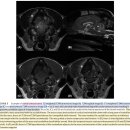
as low T1W and T2W signals resulting in cortical thickening or effacement of normal bone marrow Amorphous, sunburst, spiculated, and palisading periosteal proliferation was considered aggressive. Smooth and lamellar periosteal...
-
유방암 2015.04.11해당카페글 미리보기
현저히 떨어진다. e. screening mammography상 이상 있는 경우 - 이상 소견 : 군집성의 미세석회화, 고밀도(특히, spiculated), 구조의 뒤틀림 등 - screening mammography상 이상이 있으면 spot magnificaiton, oblique view, 흡인, 유방 초음파 등을...
-
아버지께서(월남참전) 폐암 판정을 받았는데 혜택을 볼수 있는지요? 2005.07.02해당카페글 미리보기
흉부 대동맥 invasion bronchus 기관지 invasion pleura 늑막 the enhanced ct scan of the chest reveals about 4cm sized spiculated margined soft tissue mass at left lower lobe with abuted to descending thoracic aorta and left main bronchus...
-
3월 셋째 주 reading materials 2022.03.18해당카페글 미리보기
soft tissue lesion 사이의 연관성을 찾는 것이 목적인 논문으로, CT 상 흔한 특징으로 주로 폐 후엽에서 발견되는 점, spiculated lesion margin을 보이며 내부로 air bronchogram에 확인되는 점, pleural tags, heterogeneous한 조영 증강, 주변 실질의...


