카페검색 본문
카페글 본문
-
hcc ? - > hemangioma 2026.05.09해당카페글 미리보기
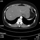
간문맥 혹은 간정맥혈전( ), 담낭이상(직접기술), 기타( 간우엽 내 석회화 ) 1. 지방간, 경도 2. 간우엽 내 S8, 1.07cm hyperechoic lesion with halo sign 3. 간우엽 내 석회화 4. 췌장미부는 장내 가스에 의해 관찰에 제한이 있음 rec) 간우엽 결절에...
-
SONOGRAPHIC IMAGING AND APPEARANCE OF LNs 2026.04.21해당카페글 미리보기
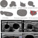
matting results in LNs unable to be distinguished from each other.11 Diffuse or focal LN echogenicity changes such as hyperechoic deposits or cystic areas within the cortex or medulla are suspicious findings.9 Reticulation (diffuse...
-
판독 form 2025.12.16해당카페글 미리보기
No significant interval change since 2023-04-19 2. Several small oil cysts in both breasts. 3. No detected previous hyperechoic mass, left breast 6 o'clock direction <Conclusion> BI-RADS Category 2 : benign finding rec) routine F/U
-
용어정리 2 2026.05.27해당카페글 미리보기
urinary tract obstruction, gas or air (diadetes mellitus) -renal fungal disease : mycetoma - fungal ball - > hyperechoic, nonshadowing, mobile structures within the renal collecting system acute : <well-defined renal pyramids> chronic...
-
경추 불안정성으로 나타나는 문제들 2024 2025.11.13해당카페글 미리보기
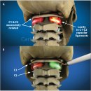
The optic nerve emerges from the posterior part of the globe and appears as a hypoechoic linear structure with a hyperechoic border (nerve sheath). The outer rim should be included in optic nerve sheath measurements and should be...
-
Re: 회전근개 석회화(극상근, 견봉하점액낭, 극하근, 견갑하근) 2018년 2025.10.16해당카페글 미리보기
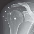
of soft tissue structures throughout the body (21, 34, 38, 74-93). On US, the calcium deposits usually appear hyperechoic with or without acoustic posterior shadowing. Bianchi and Martinoli (94) described three different calcifications...
-
Duplicated gallbladder with milk of calcium bile 칼슘 우유 담즙을 동반한 중복 담낭 2025.10.10해당카페글 미리보기
Hyperechoic fluid-without posterior acoustic shadow indicating milk of calcium bile gallbladder.
-
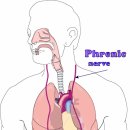
횡격막 근육의 기능부전 - 호흡불편함 phernic nerve(C3~5) 2025.08.27해당카페글 미리보기
that is increasingly being used. The diaphragm is identified ultrasonographically by a three-layer structure, with two hyperechoic outer layers representing the pleural and peritoneal membranes, and the hypoechoic layer in the middle...
-
Re : 수근관 증후군 초음파 진단!! 2026.05.22해당카페글 미리보기
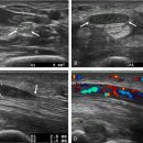
fascicle, which has a hypoechoic echo structure compared with the peri- and epineural connectives, which have a hyperechoic appearance and which are confused with the fibro-adipose connective that surrounds the nerve and the adjacent...
-
Re: 신경차단술을 위한 초음파 진단 2021 논문!! 2025.07.14해당카페글 미리보기
Center for Medical Art & Photography) Full size image The bony structures of the lumbar vertebrae appear as hyperechoic white lines on ultrasound imaging with black acoustic shadowing underneath. Figure 2 shows the bony windows through...